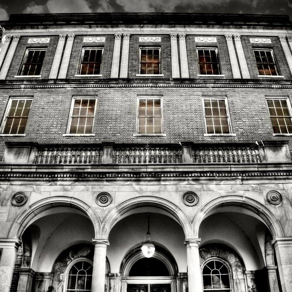
This Stanislas Keenan guy was a mover and shaker in Eloise’s history. 45 years his career spanned. He was a bookkeeper, historian of the place,Postmaster, representative for Michigan Central Railway and American Express Company. On top of that he seemed to lead the place into the high tech era of Xrays. Keenan reported in his 1913 book, “The History of Eloise,” that in the first few years people in the surrounding area heard “the chained unfortunates roaring and shrieking in discord with the squealing pigs beneath.”
Keenan, who was also an amatuerelectrical experimenter, loaned hisinduction coils and static machine to the Hospital to produce x-rays by the method described by Professor Roentgen. The Hospital, in turn, requested that he build a larger static machine; and Mr. Keenan then contructed a twelve-plate, sectorless Wimhurst machine which was finished in December, 1896, and placed in the rear of the County House dispensary. The big machine took all the space, and the Board ordered the small Hospital unit in the west wing vacated for the machine. Thus, the nucleus of one of the best and, for years, only hospital electrical laboratory in the west was established. At first the big static machine was operated by hand; later, a three-horsepower motor was purchased for it and was adapted so that single two- and three-phase currents could be taken from it. Later, a 2 1/2- kilowatt, 20,000-volt Kuhlman transformer was installed.

In the same year as Professor Roentgen’s announcment of his discovery, the Chief Bookkeeper of Eloise, Stanislas Keenan, who was also an amatuerelectrical experimenter, loaned hisinduction coils and static machine to the Hospital to produce x-rays by the method described by Professor Roentgen. The Hospital, in turn, requested that he build a larger static machine; and Mr. Keenan then contructed a twelve-plate, sectorless Wimhurst machine which was finished in December, 1896, and placed in the rear of the County House dispensary. The big machine took all the space, and the Board ordered the small Hospital unit in the west wing vacated for the machine. Thus, the nucleus of one of the best and, for years, only hospital electrical laboratory in the west was established. At first the big static machine was operated by hand; later, a three-horsepower motor was purchased for it and was adapted so that single two- and three-phase currents could be taken from it. Later, a 2 1/2- kilowatt, 20,000-volt Kuhlman transformer was installed.
The members of the Wayne County Medical Society visited the Institution soon after the installation of the first equipment; and immediately, Detroit physicians began sending patients to the Hospital laboratory for pictures of bone fractures and dislocations.
Thus, the Institution was among the first, if not the first, medical facilities in the United States to render radiographs for medical diagnosis.
This free service continued until 1900 when the volume became too great. Many of the original x-ray tubes dating from 1897 to 1917 have been preserved by the Institution and were on display in the lobby of the General Hospital. Also on display were the original Wimhurst static machine and the motor later used to power it.
Gradually, the induction coil supplanted the static machine; and high frequency came into favor. As a result, high-frequency coils of great power were installed in the laboratory, including new induction coils, operated by electrolytic circuit breakers; and the newly developed Crookes tubes were purchased as well. There still was considerable crudeness both in the apparatus, and soon alnernating current came into favor with the advent of what was commonly called the “interrupterless transformer.” This was a step-up transformer of exceedingly high voltage and heavy ampherage, utilizing a motor-driven rectifier which picked off the peaks of the alernating current wavess from the secondary of the transformer and commuted them into uni-directional pulsating current. The electrical laboratory was enlarged to include five rooms, with the retention of the original laboratory area for the generating apparatus. About this time, a newer version of the Coolidge tube supplanted the earlier types of the Crookes tubes.
The original laboratory was set up for exhibit in 1934 in four clinic rooms in the basement of “A” building. In 1936, when the Board decided to install a bateriological laboratory in that location, the electrical laboratory was dismantled; and its apparatus was stored.

It was noted that, during operation, there were burning effects of x-rays which caused acute dermatitis and in many cases which caused fatal injuries. At first it was thought the burning effect was caused by the electrical apparatus; but this was soon disproved by several scientists., especially Professor Elihu Thompson, of the General Electric Company, who showed beyond doubt that the injurious effects were caysed by the radiation. Investigation showed that the rays had a beneficial action in treating superficial skin diseases; further investigation showed that the Crookes tube of low vacuum, or “soft tubes as they were called, were more efficient that those of high exhaustion; and tube makers soon placed these on the market. As research went on, it was found that the most effective results were obtained by the use of very large high-voltage tubes of air-cooled or water-cooled targets.

In 1927 the Hospital purchased a Wappler Deep Therapy outfit, which for a time answered the purpose very well. It was of a water-cooled type and worked under a tube-circuit pressure of 18,000 to 200,000 volts and 10 to 25 milliampheres of current; it rectified the entire wave and gave very good results. But after the William J. Seymour Hospital was opened in 1933, the call for deep therapy was greatly increased; and the Hospital was unable to treat all the patients requiring deep radiation. Consequently, the old Wappler machine was dismantled; the rooms were re-altered; and a new machine was purchased which doubled the capacity. This machine, produced by General Electric, consisted of a high-tension transformer which delivered 200,000 volts and 25 milliampheres in the tube circuit. The Coolidge tube was entirely immersed in oil. This apparatus had many advantages, the principals of which were the safety features, far greater flexibility, and a greater current capacity. The greater current capacity resulted in a shorter treatment time, which consequently allowed for the handling of a greater number of patients; and the use of a thicker filter gave more homogeneous ray of shorter wavelengths which had less destructive action on normaltissue. Also, the greater flexibility of the unit allowed for treatment innearly any concievable position.
By 1929, the trochoscope which was installed in 1915 and the Bucky diaphragm system which was installed in 1921 were not taking pictures satisfactory with the tube postitioned beneath the table. In addition, this arrangement sometimes severely shocked the operator. Both were discarded in 1929 in favor of a new type of diagnostic table which eliminated these hazards. All the other x-ray apparatuses installed and used from 1915 to 1929 were dismantledand stored in January, 1933.
When the Dr. William J. Seymour Hospital, the first General Hospital of the Institution, opened in 1933, the X-ray Department was completely refurbished and placed on the fourth floor of the building. Through the ensuing years, the equipment had been constantly added to and modernized. In 1962 the new General Hospital building was opened, and all the equipment was replaced. In 1965 a Radioactive Cobalt Unit was placed in service. In 1982,a head “CAT” scanner (Computerized Axial Tomography) was installed.
On January 17th, 1950, the Detroit News Daily newspaper paid tribute and credit to Mr. Keenan for the creation of the first x-ray machine in the United States.
[ This information presented in whole from “A History of the Wayne County Infirmary, Psychiatric, and General Hospital Complex at Eloise, Michigan” by Alvin C. Clark; pages 13-16. ]
